Ultrasound-Guided Injections for Musculoskeletal Pain
Written by YiQiu Hu, NMD
Arizona-licensed naturopathic physician and virtual concierge medicine provider
Published August 2026 | Medically reviewed August 2026
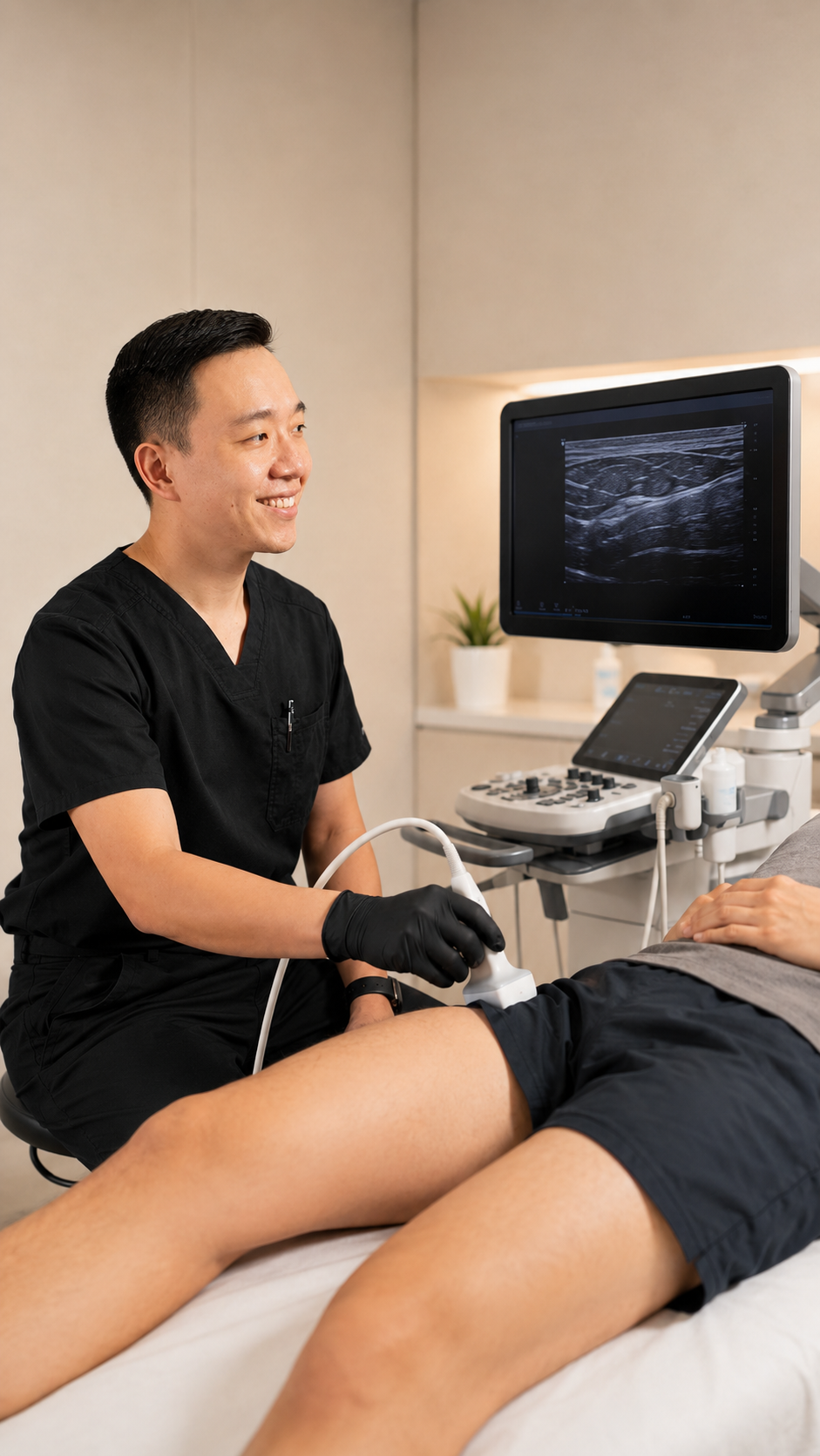
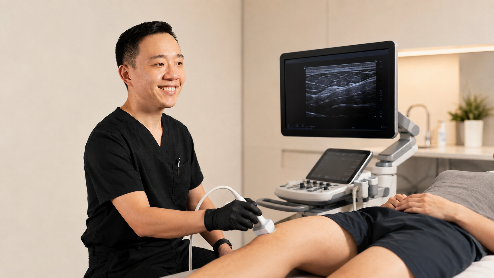
Musculoskeletal injections can be useful for selected joint, tendon, bursa, soft-tissue, and nerve-related conditions. But performing an injection involves more than simply placing a needle near the area that hurts.
The physician must determine which anatomical structure is most likely contributing to the symptoms, whether an injection is appropriate, what substance—if any—should be injected, and where the needle should be placed.
Musculoskeletal ultrasound can help with the procedural part of that process by allowing the physician to see many soft-tissue structures and monitor the needle in real time.
Research consistently shows that ultrasound guidance can improve the accuracy of needle placement for many musculoskeletal procedures. Whether that increased accuracy produces meaningfully better pain relief or function varies according to the body region, diagnosis, injection technique, and treatment being performed.
For that reason, ultrasound is best viewed as a precision tool within a broader integrative pain-management strategy—not as a treatment by itself.
What Is an Ultrasound-Guided Injection?
An ultrasound-guided injection is a procedure in which the physician uses real-time ultrasound imaging to visualize anatomical structures while advancing a needle toward a specific target.
Musculoskeletal ultrasound can display many structures involved in pain and movement, including:
Muscles
Tendons
Ligaments
Joints
Bursae
Peripheral nerves
Blood vessels
Fluid collections
Certain areas of inflammation or tissue abnormality
The ultrasound probe sends sound waves into the body and converts the returning signals into images displayed on a screen.
Unlike X-ray and CT imaging, diagnostic ultrasound does not use ionizing radiation. It also provides real-time imaging, making it particularly useful for certain needle-guided procedures.
During an injection, the physician can often observe the needle as it advances through the tissue and confirm its relationship to the intended target and surrounding anatomy.
Why Does Injection Accuracy Matter?
The painful area a patient identifies is not necessarily the exact structure that needs treatment.
For example, shoulder pain could potentially originate from the glenohumeral joint, acromioclavicular joint, subacromial bursa, rotator cuff tendons, biceps tendon, cervical spine, peripheral nerves, or other structures.
Similarly, pain near the hip might arise from the hip joint, tendons, bursae, muscles, referred lumbar pain, or surrounding nerves.
This is why a thoughtful evaluation should come before the procedure.
Our complete guide to integrative pain management explains in greater detail why identifying the probable source of pain is an important part of choosing an appropriate treatment.
Once a target has been selected, accurate needle placement can help ensure that an injection reaches the intended anatomical location while minimizing unnecessary contact with surrounding tissues.
What Does the Research Show About Ultrasound Guidance?
One of the most consistent findings in the research literature is that ultrasound guidance improves injection accuracy for many musculoskeletal targets.
For example, systematic reviews comparing ultrasound-guided and landmark-guided knee injections have reported higher placement accuracy with ultrasound. Reviews involving shoulder and other musculoskeletal procedures have shown similar trends.
The more important clinical question, however, is whether greater accuracy always results in greater pain relief.
The answer is less straightforward.
A 2024 umbrella review examining systematic reviews and meta-analyses found that ultrasound-guided musculoskeletal injections were generally more accurate than landmark-guided injections, particularly in the shoulder. However, improvements in pain and functional outcomes were less consistent across studies.
This distinction is important.
More accurate placement does not automatically guarantee a better clinical result.
Pain outcomes also depend on factors such as:
Whether the diagnosis is correct
Whether the selected treatment is effective for that diagnosis
Severity and duration of the condition
Structural tissue damage
Previous injuries or surgery
The medication or solution being injected
Rehabilitation and activity after the procedure
The patient's overall health
Individual biological response
The European Alliance of Associations for Rheumatology has similarly noted that imaging guidance such as ultrasound may be used to improve the accuracy of intra-articular therapy when available.
Which Musculoskeletal Injections Can Be Performed With Ultrasound Guidance?
Ultrasound guidance may be useful for a wide range of procedures, depending on the diagnosis and anatomical location.
Joint Injections
Ultrasound can help guide injections into selected joints, such as the:
Shoulder
Hip
Knee
Elbow
Wrist
Ankle
Small joints of the hands or feet
Joint injections may be performed for different reasons depending on the condition, including selected cases of osteoarthritis, inflammatory joint conditions, or diagnostic evaluation.
The decision to inject a joint should depend on the diagnosis and overall treatment plan rather than the presence of pain alone.
Bursa Injections
Bursae are small fluid-containing structures that reduce friction between tissues.
Irritation of a bursa may contribute to pain in areas such as the shoulder, hip, elbow, or knee.
Ultrasound can help a physician identify the bursa and guide a needle into the intended space when an injection is appropriate.
Tendon and Tendon-Sheath Procedures
Certain tendon disorders may involve the tendon itself, its surrounding sheath, or adjacent tissue.
Ultrasound can help distinguish these structures and guide treatment to a specific location.
This precision is especially important because the intended target depends on the diagnosis and injectate. Some treatments are designed to be placed around a tendon rather than directly into tendon fibers.
Peripheral Nerve Procedures
Many superficial peripheral nerves can be visualized with musculoskeletal ultrasound.
This can help physicians perform selected nerve blocks, hydrodissection procedures, or other perineural treatments while visualizing the nerve and nearby blood vessels.
These procedures require careful diagnosis because numbness, tingling, burning, or radiating pain can have multiple causes, including problems that originate farther upstream in the spine or nervous system.
Aspiration of Fluid
Ultrasound may also help guide aspiration when excess fluid is present within a joint, bursa, cyst, or other accessible structure.
In some cases, removing fluid can help relieve pressure or provide a sample for diagnostic testing.
A suspected infected joint or other serious condition requires appropriate medical evaluation rather than routine injection treatment.
What Can Be Injected?
Ultrasound describes how the needle is guided, not what is injected.
Different procedures may involve different substances depending on the diagnosis.
Examples may include:
Corticosteroids
Corticosteroid injections are commonly used for selected inflammatory joint and soft-tissue conditions.
They may provide short-term pain relief in certain conditions, but they are not appropriate for every patient or every musculoskeletal problem. The potential benefits of repeated corticosteroid injections must also be weighed against their risks.
Local Anesthetics
Local anesthetics may sometimes be used to reduce procedural discomfort or as part of a diagnostic or therapeutic injection.
Temporary improvement after an accurately targeted anesthetic injection can occasionally provide useful information about whether a particular structure is contributing to symptoms.
Platelet-Rich Plasma
Platelet-rich plasma, or PRP, is prepared from a patient's own blood and contains a concentrated platelet fraction.
PRP is being studied and used for selected tendon, ligament, and joint conditions, but outcomes and the quality of evidence vary significantly according to the condition and preparation technique.
Ultrasound guidance may be used to place PRP into or around a selected anatomical target when the procedure is appropriate.
Dextrose-Based Treatments
Dextrose solutions may be used in certain prolotherapy or perineural injection techniques.
As with other injection therapies, evidence differs considerably depending on the diagnosis and technique.
Patients interested in these treatments can explore additional physician-written information in our integrative pain management resource center.
When Is Ultrasound Guidance Particularly Helpful?
Not every musculoskeletal injection requires imaging guidance.
Some superficial anatomical targets can be injected reliably using established landmarks, particularly when the anatomy is easy to identify and the clinician has extensive experience with the technique.
Ultrasound guidance may become especially useful when:
The target is small
The target is located near nerves or blood vessels
The patient's anatomy is difficult to identify by palpation
Previous surgery has altered the anatomy
A joint has significant degenerative changes
A tendon sheath or specific soft-tissue plane needs to be reached
Fluid needs to be aspirated
A previous landmark-guided procedure was unsuccessful
Precise placement of a higher-cost treatment such as PRP is desired
The goal is not to use technology simply because it is available. The goal is to select the safest and most appropriate technique for the particular procedure.
Can Ultrasound Help Avoid Nearby Nerves and Blood Vessels?
One practical advantage of ultrasound is that the physician can often visualize structures surrounding the intended injection target.
Blood vessels can frequently be identified with Doppler imaging, while many peripheral nerves can be recognized based on their location and ultrasound appearance.
This information may help the physician choose a needle path that avoids structures that should not be entered.
Ultrasound nevertheless does not eliminate procedural risk. Safe injections still require appropriate training, knowledge of anatomy, sterile technique, patient selection, and careful needle control.
Does Ultrasound Make an Injection Safer?
Ultrasound guidance can improve visualization and needle-placement accuracy, but no injection is completely risk-free.
Potential complications depend on the treatment and anatomical location and may include:
Temporary procedural pain
Post-injection soreness
Bruising
Bleeding
Infection
Allergic reaction
Temporary numbness or weakness
Injury to a nerve or blood vessel
Medication-specific adverse effects
Failure to improve symptoms
There may also be risks specific to corticosteroids, local anesthetics, PRP, dextrose, or another substance being used.
Before a procedure, patients should tell their physician about relevant medical conditions, allergies, current infections, bleeding disorders, anticoagulant or antiplatelet medications, previous reactions to injections, and other important health information.
Medication-specific considerations may also need to be reviewed.
What Happens During an Ultrasound-Guided Injection?
The exact process varies according to the procedure, but many outpatient ultrasound-guided injections follow similar steps.
First, the physician reviews the diagnosis, treatment goals, potential benefits, risks, alternatives, and relevant medical history.
The patient is then positioned so the target area can be safely accessed.
Ultrasound gel is placed on the skin, and the physician uses the ultrasound transducer to identify the target and nearby anatomical structures.
The skin is cleaned using an appropriate antiseptic technique.
The needle is then advanced while the physician monitors its position on the ultrasound screen. Depending on the technique, the needle may be visualized along much of its path or monitored as it approaches the intended structure.
Once placement is confirmed, the selected medication or solution is administered.
The needle is removed, and a small bandage may be placed over the injection site.
The entire process is often completed during an outpatient visit, although preparation and recovery vary according to the specific procedure.
What Should Patients Expect After an Injection?
Post-procedure recommendations depend heavily on what was injected and which structure was treated.
Some patients return to normal daily activity relatively quickly, while others may be advised to temporarily limit strenuous exercise or loading of the treated tissue.
Mild soreness may occur after many injection procedures.
Patients should receive individualized instructions explaining:
Expected symptoms
Activity restrictions
When to resume exercise
Whether rehabilitation should continue
When improvement may occur
Which symptoms require medical attention
When follow-up should occur
For a broader overview of how injections fit into pain treatment, read A Patient's Guide to Starting Integrative Pain Management.
Ultrasound Guidance Does Not Replace an Accurate Diagnosis
Perhaps the most important limitation of ultrasound-guided procedures is that perfect needle placement cannot compensate for treating the wrong problem.
A technically accurate injection into the shoulder joint will not solve pain caused primarily by cervical radiculopathy.
A precisely placed knee injection will not correct certain mechanical injuries requiring orthopedic management.
Similarly, pain that is primarily related to nerve sensitization, widespread pain mechanisms, referred pain, systemic inflammatory disease, infection, fracture, or another medical condition may require a very different treatment strategy.
This is why injection therapy should begin with a clinical question:
What structure or condition are we trying to treat, and why do we believe this procedure will help?
Ultrasound then becomes one tool for carrying out that treatment plan more precisely.
Ultrasound Is Also a Diagnostic Tool
Musculoskeletal ultrasound can sometimes be used during the same clinical evaluation to assess structures such as tendons, muscles, ligaments, joints, nerves, and fluid collections.
This can be especially useful because ultrasound is dynamic. The physician may observe certain structures while the patient moves rather than viewing anatomy only in a static position.
However, ultrasound does not replace every other imaging modality.
X-rays may be more useful for evaluating certain bone and joint changes. MRI may provide more comprehensive information for some deep structures, bone-marrow abnormalities, complex internal joint pathology, or conditions not adequately visualized with ultrasound.
The correct imaging test depends on the clinical question.
Where Do Ultrasound-Guided Injections Fit Into Pain Management?
An injection should rarely be viewed as the entire treatment plan.
Depending on the condition, a comprehensive strategy may also involve:
Activity modification
Progressive therapeutic exercise
Physical therapy
Strengthening
Mobility work
Weight management
Ergonomic changes
Sleep optimization
Appropriate medications
Acupuncture or dry needling
Bracing or supportive devices
Regenerative medicine
Specialist referral
Surgical evaluation when necessary
For some patients, an injection may reduce symptoms sufficiently to make rehabilitation easier.
For others, the procedure may have little benefit, prompting reassessment of the diagnosis or treatment strategy.
Follow-up is therefore important.
Instead of judging success only by a pain score, physicians may also evaluate whether the patient can walk farther, sleep better, return to exercise, regain strength, perform occupational tasks, or participate more comfortably in daily activities.
Are Ultrasound-Guided Injections Better Than Landmark-Guided Injections?
The most accurate answer is: sometimes, depending on what outcome is being measured.
For needle placement, the evidence generally favors ultrasound guidance for many anatomical targets.
For pain relief and functional improvement, the advantage is less universal. Some studies show improved outcomes with ultrasound guidance, while others find similar results when an experienced clinician performs a landmark-guided procedure correctly.
Therefore, ultrasound guidance should not be marketed as automatically superior in every situation.
Its most defensible advantages are the ability to visualize anatomy, observe needle placement in real time, improve accuracy for many targets, and help the clinician plan the needle path around surrounding structures.
A Personalized Approach to Musculoskeletal Injections
At Ask Dr. Hu, injection procedures are considered within a broader integrative pain-management evaluation.
Not every painful condition requires an injection, and not every patient is an appropriate candidate for a particular procedure.
Treatment selection may depend on:
Medical history
Physical examination
Probable diagnosis
Previous imaging
Diagnostic ultrasound findings when appropriate
Treatments already attempted
Functional limitations
Patient preferences
Potential risks and benefits
Rehabilitation needs
Whether additional imaging or specialist referral is necessary
The objective is not simply to perform a procedure. It is to determine whether a procedure makes sense within the patient's overall treatment plan.
The Bottom Line
Ultrasound-guided injections allow physicians to visualize many musculoskeletal structures and monitor needle placement in real time.
Research shows that ultrasound guidance can improve procedural accuracy for many joint and soft-tissue injections. Whether it produces substantially better pain relief or functional outcomes than a well-performed landmark-guided injection varies by diagnosis and anatomical location.
The quality of the overall decision-making remains just as important as the technology.
A successful procedure begins with an appropriate diagnosis, selection of the correct treatment and anatomical target, careful procedural technique, and a plan for rehabilitation and follow-up.
If you are experiencing persistent joint, tendon, muscle, or other musculoskeletal pain, you can learn more about integrative pain management at Ask Dr. Hu or contact our office to discuss whether an evaluation may be appropriate.
Medical Review & Editorial Standards
This physician article was written for educational purposes by an Arizona-licensed naturopathic physician with clinical experience in musculoskeletal evaluation, integrative pain management, diagnostic ultrasound, and injection-based treatment approaches.
The discussion of ultrasound guidance reflects current medical literature showing that image guidance can improve injection accuracy for many musculoskeletal procedures while recognizing that improvements in clinical outcomes vary by condition.
Key references used in preparing and medically reviewing this article include the European Alliance of Associations for Rheumatology recommendations for intra-articular therapies, the 2024 umbrella review of ultrasound-guided versus landmark-guided musculoskeletal injections, and the U.S. Food and Drug Administration's information on diagnostic ultrasound imaging.
Treatment recommendations must be individualized. The appropriateness, risks, benefits, and evidence supporting a particular injection depend on the diagnosis, anatomical location, medication or solution being used, the patient's medical history, and available alternatives.
Last medically reviewed: August 2026
About the Author
YiQiu Hu, NMD
Dr. YiQiu Hu is an Arizona-licensed naturopathic physician and founder of Ask Dr. Hu. His clinical work includes concierge medicine, preventive healthcare, health optimization, laboratory evaluation, medical weight management, hormone care, and integrative pain management.
His approach to musculoskeletal pain emphasizes careful evaluation, identification of the likely pain-generating structures, discussion of conservative and procedural options, and individualized treatment planning rather than relying on a single therapy for every patient.
Dr. Hu earned his Doctor of Naturopathic Medicine degree from Sonoran University of Health Sciences and a Bachelor of Science in Biomedical Sciences from Northern Arizona University. He also has experience in medical leadership and multidisciplinary healthcare practice.
Learn more about Dr. YiQiu Hu and the physicians at Ask Dr. Hu.
This article is intended for general educational purposes only and does not replace individualized medical advice, diagnosis, or treatment. Reading this article does not establish a physician-patient relationship. If you are experiencing a medical emergency, call 911 or seek appropriate emergency medical care.